Echocardiogram (ECHO)
Echocardiography is a test that creates live images of your heart using sound waves. The image is called an echocardiogram. This test allows your doctor to monitor how well your heart and its valves are working.
It can detect blood clots in the heart chambers, fluid in the sac around the heart, issues with the aorta, problems with the pumping or relaxing function of the heart, valve dysfunction, and pressure abnormalities inside the heart.
An echocardiogram is key in determining the health of the heart muscle, especially after a heart attack. It can also detect heart defects or irregularities in the fetus.
Dr. Vivek Kumar is the best Echocardiography Doctor in Ghaziabad, Delhi NCR and Director – Interventional Cardiology & Head of Structural Heart Program at Max Super Speciality Hospital, Vaishali. An echocardiogram is a painless procedure with extremely rare risks.
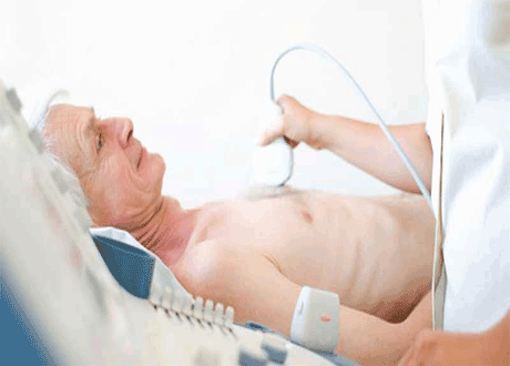
Echocardiogram (ECHO) in Ghaziabad, Delhi NCR
Types of Echocardiograms
- Transthoracic Echocardiogram (TTE): The most common type. A transducer is moved over the chest to capture images of the heart.
- Transesophageal Echocardiogram (TEE): A probe is passed down the esophagus for clearer, more detailed images of the heart structures.
- Stress Echocardiogram: Images are taken before and after exercise or medication that increases heart rate to assess how the heart responds to stress.
- 3D Echocardiography: Creates three-dimensional images of the heart, useful for diagnosing complex problems and planning valve surgery.
- Fetal Echocardiography: Safe ultrasound test performed during pregnancy to check the unborn baby’s heart for defects.
Echocardiography under Stress
In a stress echocardiogram, transthoracic images are taken before and after exercise or medication that makes the heart beat faster. This helps the doctor assess how the heart responds to physical stress and detect reduced blood flow that may not appear at rest.
What to Expect
Patches connected to the echocardiogram machine will be applied to your chest. The machine and other devices collect data at regular intervals to evaluate heart function.
They will measure your:
- Heartbeat, breathing, and blood pressure
- Response during an exercise stress test (come prepared to work out)
- A contrast dye may be injected to obtain a clearer image
- Heart rate and blood pressure before, during, and after exercise
3D Echocardiography
A three-dimensional (3-D) echocardiogram creates a detailed 3-D image of your heart using multiple views from transthoracic or transesophageal echocardiography. It is especially useful for diagnosing heart problems in children and for planning heart valve surgery.
Echocardiography during Pregnancy (Fetal Echo)
The transducer is placed over the pregnant woman’s abdomen to check for heart problems in the fetus. The test is considered completely safe for the unborn child because, unlike an X-ray, it does not use radiation.
Risks
- Echocardiograms are considered very safe. Unlike X-rays, they do not use radiation.
- There is a slight risk of allergic reaction if contrast dye or agitated saline is used. Contrast should never be used during pregnancy.
- Mild skin discomfort may occur when EKG electrodes are removed.
- Transesophageal echocardiogram carries a small risk of throat irritation or, very rarely, esophageal injury.
- The most common side effect after TEE is a sore throat. Sedation may cause temporary drowsiness.
- Stress echocardiogram (exercise or medication) carries a very small risk of irregular heartbeat or, rarely, heart attack.
During the Procedure
- Most echocardiograms are performed in a hospital or clinic and take less than an hour.
- Transthoracic Echo: You undress from the waist up. Electrodes are attached. The technician moves a transducer across your chest while you may be asked to breathe or change position.
- Transesophageal Echo: Your throat is numbed and a mild sedative is given. A thin tube with a transducer is guided down the esophagus to obtain clear images.
- Stress Echo: Similar to transthoracic echo, but images are taken before and after 6–10 minutes of exercise (or medication).
Preparing for an Echocardiogram
There is usually no preparation needed for a standard transthoracic echocardiogram.
For a transesophageal echocardiogram, you will be asked not to eat or drink for about 8 hours before the test. You may also be advised not to drive for a few hours afterward because of sedation.
Wear comfortable exercise clothes and shoes if a stress echocardiogram is planned.
Recovery
- Most echocardiograms require little to no recovery time.
- After a transesophageal echocardiogram you may have mild throat soreness for a few hours.
- You can usually resume normal activities the next day.
What the Results Can Show
An echocardiogram can reveal abnormal chamber size, pumping function problems, heart muscle stiffness, valve issues, blood clots, reduced blood flow during exercise, and elevated pressures inside the heart.
If any abnormality is found, Dr. Vivek Kumar will discuss the findings with you and recommend the most appropriate treatment plan, which may include further tests or specialist care.
Takeaway
- Echocardiograms provide accurate, real-time information about how well your heart is working and help identify areas of concern.
- Most procedures are non-invasive. Contrast or TEE may be used when clearer images are needed.
- Echocardiography is one of the safest and most reliable tools for diagnosing heart conditions and guiding treatment.
- For expert echocardiography in Ghaziabad and Delhi NCR, consult Dr. Vivek Kumar at Max Super Speciality Hospital, Vaishali.